Subscriber Content
Add content here that will only be visible to your subscribers.

Rods and cones are photoreceptors present in the -retina. Each rod and cone are divided into 1.Outer segment 2.Inner segment, and 3.Synaptic zone.
In the rods, the discs are separated from the cell membrane. However, saccules in the cones are formed by the infolding of the cell membrane.
The outer segment of the rod is a thin rod-like structure, while that of the cone is conical.
The photosensitive pigment in the rod is rhodopsin, which is very light-sensitive. This rhodopsin is responsible for dim light vision, i.e., scotopic vision.
In a cone, the photosensitive pigment is idopsin, responsible for bright light vision, photopic vision, color, and high visual acuity.
Rods are abundant in the human eye. It is estimated that about 120 million and 6 million cones are in each eye.
Rods are abundant in the peripheral parts of the retina and absent in the fovea. Fovea has only cones.
In rods, the photosensitive pigment is rhodopsin, which is present in the discs. It is coupled to the G-protein and is known as Visual purple.it is purple-red and stimulated by blue-green light with a wavelength of 505nm.
In cones, the photosensitive pigment is idopsin in the saccules. Structures of rhodopsin and idopsin are the same, except the latter has “photopsin” instead of “scotopin.”
The rhodopsin is composed of 1.retinene1 and scotopin.
Retinene1 is an aldehyde of vitaminA1.
In the dark, retinene is in the 11 cis-form. When light strikes, rhodopsin 11 cis-form is converted into trans-form, and scotopic is released. Rhodopsin gets bleached.
1. Some bleached rhodopsin recover directly.
2. The ‘alcohol dehydrogenase’ enzyme reduces the majority of trans-retinene in the presence of NADH to vitamin A1. This, in turn, combines with scotopin to form rhodopsin.
Cones are divided into three types according to the presence of pigments in their saccules.
1. Cyanolabe responds maximally to blue light, with a wavelength of 430 nm.
2. Chlorolabe – responds maximally to green light, with a wavelength of 535 nm.
3. Erythrolabe- responds maximally to red light, with a wavelength of 575 nm.
These three pigments are responsible for the primary colors: blue, green, and red. Other colors are a mixture of these colors in different proportions.
Humans can see light of wavelength between 400 nm to 750 nm.
The choroid layer of the retina absorbs ultraviolet light (wavelength of less than 400 nm).
Infrared light has a wavelength above 750 nm, and the cornea absorbs it.
The visual threshold is the minimum amount of light that produces a sensation of light.
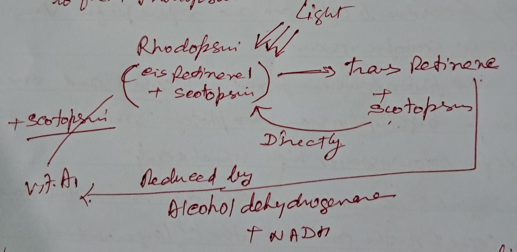
Effect of light on rhodopsin and its regeneration.
Cones are responsible for bright light vision, color vision, and excellent visual acuity.
Between 0.001 to 1 m. A is a transitional zone called the “Duplicity Theory of Vision.”
Below 0.1 m A, the reading is complex, and prolonged exposure to light 100000 m A will damage the retina.
Add content here that will only be visible to your subscribers.
Read more of this content when you subscribe today.