Functions and Structure of the Uterus Explained
Introduction
The uterus is a hollow muscular organ situated in the female pelvis. It is a pear-shaped (Pyriform) organ that lies between the urinary bladder in front and the rectum behind. The uterus is usually anteflexed and anteverted.
The word uterus is derived from Latin uterus or womb. The uterus is a hormone-sensitive female sex organ that contains muscles and glands in the endometrium.
The size of the uterus in an adult female is 3 inches x 2 inches x 1 inch (7.5 cm long x 5 cm wide x 2.5 cm thick. The weight of the adult nulliparous uterus ranges from 50 to 80 g.
The uterus develops from the fused vertical part of the two Müllerian ducts.
Table of Contents
- Relations
- Parts of the uterus
- Histology
- Secretion of the uterus
- Arterial supply
- Veins
- Lymphatic:
- Nerve supply:
- Support of the uterus
- Functions of the uterus
Relations
Anteriorly, the body forms the posterior wall of the uterovesical pouch and below the base of the bladder. The anterior surface of the body is also known as the vesical surface. It is flat and related to the urinary bladder, from which it is separated by a peritoneal fold, forming the posterior and upper part of the vesicouterine pouch. The vesicouterine pouch is also known as the uterovesical pouch.
The posterior surface of the body of the uterus is known as the intestinal surface. It is convex. The peritoneum separates the posterior wall of the body of the uterus from the coils of the terminal ileum and the sigmoid colon. Thus, it forms the anterior wall of the pouch of Douglas or rectouterine pouch.
Lateral borders are rounded and convex. The Broad ligament is attached to the lateral border, and the uterine artery is connected to the lateral border. Mackenrodt’s ligament attaches from the internal os to the supravaginal cervix and lateral vaginal wall.
Cornua of uterus; the supralateral angle of the body at the junction of the body and fundus is called the cornua of uterus. Here, the uterine tubes, the ligament of the ovaries, and the round ligament of the uterus are attached.
It opens at the upper end on both sides to the uterine tubes, and it is continuous with the Vagina.
Parts of the uterus
1. Body or corpus
2. Isthumus
3. Cervix

Parts of the Uterus
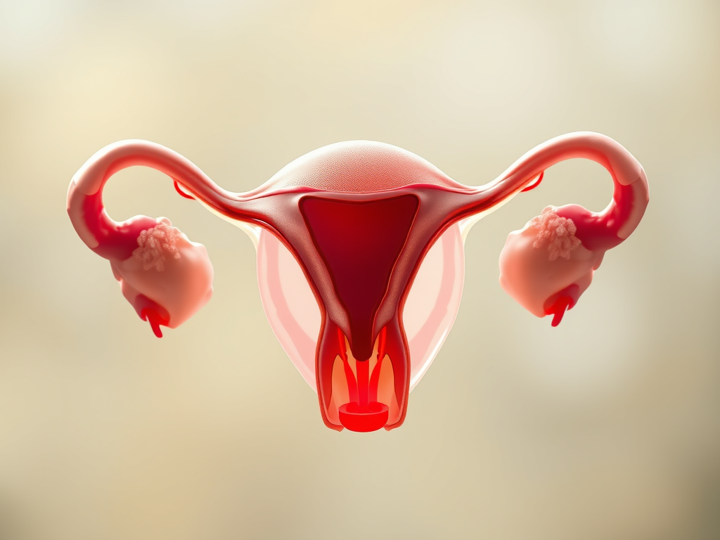
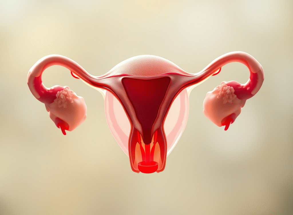
The Uterus and its parts. Credit to canva
The body or corpus is divided into
Fundus –the part of the uterus which lies above the uterine tubes. It is convex, has a free upper end, and is covered with the peritoneum. A fertilized oocyte is implanted in the posterior wall of the fundus and upper part of the body of the uterus.
The body proper is the part of the uterus that lies between the fundus above and the isthmus below.
The isthmus is situated between the body and cervix of the uterus. It is a constricted part of the uterus and measures only 0.5 cm. Isthumus is limited above by the anatomical internal os and below by the histological internal os.
The cervix is cylindrical and extends from the isthmus to the external os. It has two parts: 1. The vagina part that is above the Vagina, and the other part is the vaginal part that is within the Vagina.
The cavity is triangular, with the base above and the apex below, and measures approximately 3.5 cm. It continues with the cervical canal.
Histology
The body or corpus consists of three layers.
Parametrium is the outermost serous coat that invests the entire uterus, except for the lateral border. It is adherent to the underlying muscles.
Myometrium consists of thick bundles of smooth muscle fibres held by connective tissues. The muscles are arranged in three layers: the outer longitudinal, the middle interlacing, and the inner circular.
The submucous layer is lacking.
Endometrium: The mucous layer is known as the endometrium, which is directly opposed to the muscle coat. It consists of lamina propria and surface epithelium.
The lamina propria contains stromal cells, endometrial glands, vessels, and nerves.
The surface epithelium is a single layer of ciliated columnar epithelium.
The cervix is composed mainly of fibrous tissues, with only ten to fifteen percent being smooth muscle. The vaginal part of the cervix is lined with stratified squamous epithelium.
Secretion of the uterus
The endometrial secretion is scanty and watery. Secretion of the cervical glands is alkaline and thick.
Blood supply:
Arterial supply
The main blood supply is from the uterine arteries, one on each side. The artery is a direct branch of the anterior division of the internal iliac artery.
Other sources are the ovarian and vaginal arteries with which the uterine arteries anastomose. These arteries supply
Uterus, Ovary, medial two-thirds of the uterine tube, Vagina, and structures present in the ovarian ligaments.
Veins
The veins correspond to the arteries and drain into the internal iliac veins.
Lymphatic:
Lymphatics drain into
Preaortic and lateral aortic groups – from the fundus and upper part of the body of the uterus-
Superficial inguinal gland – Cornua.
External inguinal gland- lower part of the body.
Cervix- external iliac and obturator lymph nodes
Internal iliac groups and
Sacral groups.
Nerve supply:
Sympathetic system- T5 AND T6 motor and T10 TO L1 sensory.
Parasympathetic motor and sensory by S234.
The cervix is insensitive to touch, heat, or cold.
The uterus is insensitive to handling and even to incision over its wall.
Support of the uterus
Primary
Muscular support provides active support: pelvic diaphragm, perineal body, and distal urethral sphincter.
Mechanical support fibromuscular: round ligament of uterus, Mackenrodts ligament,pubocervical ligament, uterine axis, uterocervical ligaments.
Secondary
Secondary support: by the peritoneal ligaments, the rectouterine pouch and the peritoneal fold, the vesico-uterine pouch and the fold of peritoneum.
Functions of the uterus
Accommodates and protects the fertilized ovum, and provides nutrition to the fertilized ovum
Expulsion of the foetus.
By Priyanka Kr. Checked and posted by Dr. Bijay Krishna

Please subscribe to receive the latest posts instantly. Thank you.